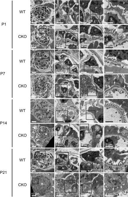
Figure 3.
The prorenin receptor-deleted podocytes developed foot process effacement and intracellular vacuoles. Electron microscopic examination of kidneys was performed from wild-type (WT) and conditional prorenin receptor-knockout (CKO) mice on postnatal day (P) 1, P7, P14, and P21. On P1, there were no obvious differences between WT and CKO mice. As early as P7, kidneys from CKO mice were found to contain prominently enlarged podocytes with extensive foot process effacement and actin filament aggregation. Numerous electron-dense autophagic vacuoles containing partially digested cellular components were observed in the cytoplasm of these podocytes. Mesangial matrix expansion was also noted in CKO glomeruli. Ultimately, on P21, the capillary lumen of kidneys from CKO mice had collapsed because of the presence of highly vacuolated podocytes and mesangial expansion. Basement membranes were intact without any electron-dense deposits in any of the glomeruli. Scale bars, 10 μm (left panels), 5 μm (left center panels), 2 μm (right center panels), and 1 μm (right panels). C, glomerular capillary; E, endothelial cells; P, podocytes; V, vesicular structure. Arrows indicate foot processes. White arrowheads indicate hypertrophic change of parietal epithelial cells. Black arrowheads indicate glomerular basement membrane. Asterisks indicate mesangial expansion and sclerosis.

