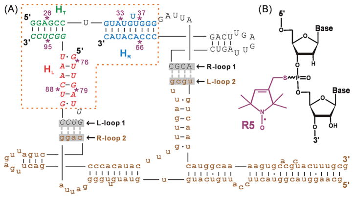
Figure 1.
(A) Dimer construct used for SDSL mapping of pRNA 3-wj global structure (see also Supporting Information Figure S1). Upper-case letters show the two respective RNA strands constituting br_B/a′, and brown lower-case letters show the unlabeled full-length monomer A/b′. The 3-wj is indicated by the dotted box, with the HT (green), HR (blue), and HL (red) helices marked. Spin labeling sites are indicated by “*” and numbered according to the corresponding full-length pRNA sites. The two sets of interacting R- and L-loops are marked and shadowed. (B) The R5 spin label. Note that following previously validated distance measurement protocols,51,70,71 all data reported here were acquired without separating the Rp and Sp phosphorothioate diastereomers present at each attachment site.

