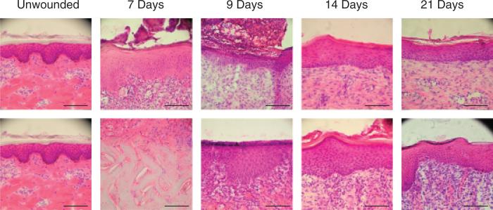
Figure 3.
Photomicrographs of 0.022 in. cut depth wounds treated with Xeroform™ (top) or semi-interpenetrating network (bottom) after postoperative days 7, 9, 14, or 21. Tissues are oriented within the photograph to align the apical surfaces of each wound section, thus focusing on the epidermis. Where epidermis has not yet formed, the interface between the underlying dermis and the fibrin clot matrix is aligned. (Hematoxylin & eosin stain, objective magnification ×20, scale bar represents 0.1 mm).

