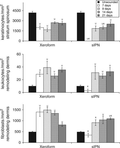
Figure 4.
Cellular density within 0.022 in. cut depth wounds dressed with Xeroform™ or semi-interpenetrating network (sIPN). Viable keratinocyte density with the stratum spinosum (top), leukocyte density within the remodeling dermis (middle), and fibroblast density with the remodeling dermis (bottom) are displayed. Data are reported as an average of 10 total viewing regions from biopsies of two separate pigs ± standard error. X represents significant difference from Xeroform™. U represents significant difference from unwounded tissue (p < 0.05).24

