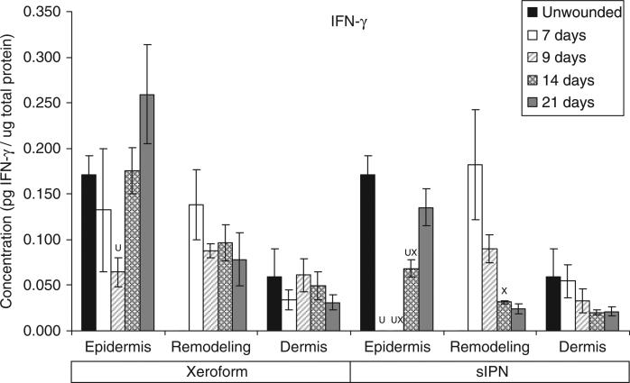
Figure 9.
IFN-γ concentration in epidermal, remodeling dermal, and dermal tissues wounded at 0.022 in. cut depth and treated with Xeroform™ or semi-interpenetrating network (sIPN). Data shown as mean ± standard error of n=2 pigs and three replicates of each n-value for each time point and treatment type. X represents significant difference from Xeroform™, and U represents significant difference from unwounded tissue at p < 0.05. Remodeling dermal tissue is statistically compared with the unwounded dermal tissue for all treatment types and time points.

