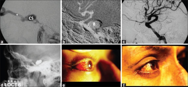
Figure 1.

A 41-year-old man with red eye, proptosis, chemosis, bruit and visual loss of the right eye (Case #1). (a) Cerebral angiography of the right internal carotid artery (ICA) confirmed high-flow direct carotid cavernous fistula with “vascular steal” phenomenon. The cavernous sinus and superior ophthalmic vein (*) showed marked dilatation. (b) Under road mapping technique, two gold-valve balloons (B) were detached. (c) Immediate angiography after balloon embolization showed complete obliteration of the fistula, preserving the ICA lumen. (d) Cranial X-ray shows contrast-filled balloons. Patient's eye (e) pre-embolization and (f) 1 week post-embolization. The patient experienced marked visual improvement
