
Figure 3.
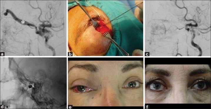
A 44-year-old female with red eye, bruit, glaucoma, and diplopia of the right eye (Case #23). (a) Cerebral angiography of the right internal carotid artery showed a CCF Type D with exclusive drainage into the superior ophthalmic vein (*). (b) A surgical exposure of the SOV was performed in order to place a microcatheter at the fistulous site of the carotid-cavernous fistula. (c) Cerebral angiography post-embolization showed complete obliteration of the fistula. (d) Cranial X-ray showed a coiled mass packed into the cavernous sinus. Patient's eye (e) pre-embolization and (f) 3 months after the endovascular procedure