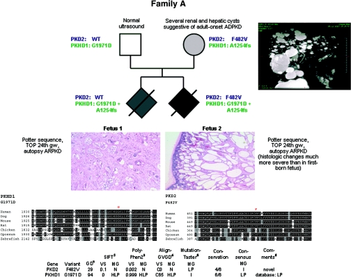
Figure 1.
Pedigree of family A with clinical information, genotypes, multisequence alignments, and bioinformatic data for both missense changes detected. Renal histology shows much more severe changes in the second-born fetus than in the first-born as regards size and number of renal cysts. (Top right panel) MR cholangiogram (T2-weighted gadolinium-enhanced coronal projection) of the mother of both fetuses in family A at 35 years of age showing multiple cysts of various sizes. The multiple hepatic cysts do not communicate with the biliary tree. Liv, Liver; R Kd, right kidney; S, superior; I, inferior; LA, left anterior; RP, right posterior.

