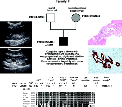
Figure 6.
Pedigree and genotypes of family F. (Left panel) Renal ultrasound of the propositus at the age of 15 years with increased echogenicity, one large cyst in the left kidney (bottom panel) and loss of corticomedullary differentiation. (Right top panel) Liver histology at the age of 9 years showing ductal plate malformation with irregularly distributed, dilated portal vein branches (asterisk), and dilated bile ducts (cross) in a fibrotic, expanded portal field (hematoxylin and eosin; original magnification, ×40). (Right bottom panel) Liver histology at the age of 15 years in line with ductal plate malformation and congenital hepatic fibrosis and demonstrating a portal tract with irregular, circular arrangement of widened bile ducts (brown) that extend into the hepatic lobules. Note absence of inflammation and fibrosis in this portal tract (Cytokeratin 7 immunoperoxidase staining; original magnification, ×200). (Bottom panel) Bioinformatic prediction scores and multiple sequence alignments obtained for the PKD1 change L2696R.

