
Figure 2.
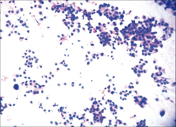
Photomicrograph showing nuclear grade II duct carcinoma. The cells are forming microacini and loose clusters. Nuclei are three to four times the erythrocytes, with granular nuclear chromatin and smooth contour. (H & E, ×100)
Official websites use .gov
A
.gov website belongs to an official
government organization in the United States.
Secure .gov websites use HTTPS
A lock (
) or https:// means you've safely
connected to the .gov website. Share sensitive
information only on official, secure websites.
Photomicrograph showing nuclear grade II duct carcinoma. The cells are forming microacini and loose clusters. Nuclei are three to four times the erythrocytes, with granular nuclear chromatin and smooth contour. (H & E, ×100)