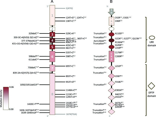
Figure 1. LGI1 mutations cluster in leucine-rich repeat (LRR) domain.
(A) LGI1 gene structure with the reported mutation sites indicated. For superscripts next to mutations, the first digit indicates the exon localization of the mutation, and the second digit (after the decimal point) indicates the order of the mutation within the exon (mutations at the same nucleotide numbered arbitrarily). i = intronic mutation; ∗ = mutation found in 2 families (n = 3); # = mutation found in a family and a sporadic case (n = 2). (B) Lgi1 protein with effect of mutations on protein summarized. The 4 LRRs in the LRR domain are represented by ovals and the 7 epitempin (EPTP) repeats in the EPTP domain are represented by diamonds. Different intensities of the red shade correspond to the density of mutations in each exon. The superscripts next to protein variants are cross-indexed to the nucleotide changes listed in the gene structure (A). CRR = cysteine-rich region; SP = signal peptide.

