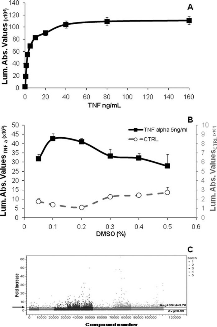
Fig. 1.
Biological behavior of stable cell line p65-NF-κB-luc. A: Concentration-dependent effect of TNF-α on Luc expression. SH-SY5Y-C1 cells were plated in 96-well plates at 20,000 cells per well and allowed to settle for 24 hr. Cells were then exposed to graded concentrations of TNF-α for 24 hr. Luc activity was measured using the Bright-Glo Luciferase assay system. B: Effect of DMSO on SH-SY5Y-C1 viability. Cells were treated with graded concentrations of DMSO in the presence (black line, left axis) and absence (gray line, right axis) of TNF-α 5 ng/ml for 24 hr. Cell viability was determined using the Cell Titer Glo assay system. C: High-throughput screening. The figure shows an example of a large library screening (112,000 compounds). The screening was completed in five independent blocks, with each block representing individual experimental days and gray coded. The data were analyzed and plotted in one graph. It was calculated that values higher than 3.78-fold the baseline value (black arrow) were statistically significant and therefore considered as hits.

