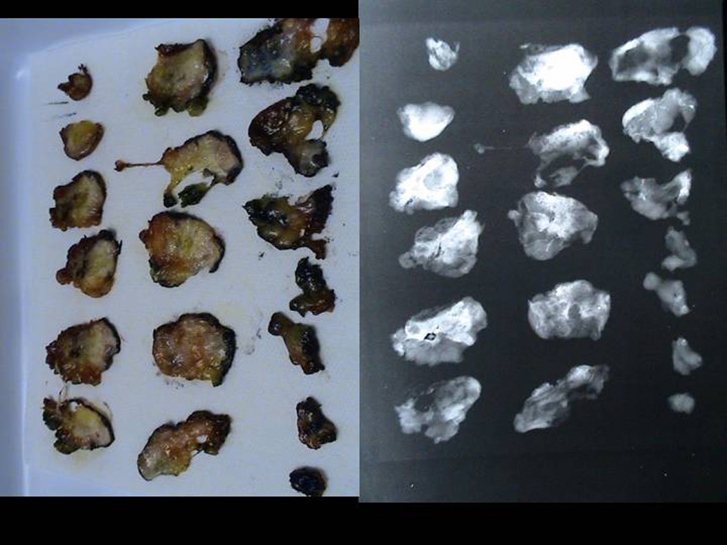
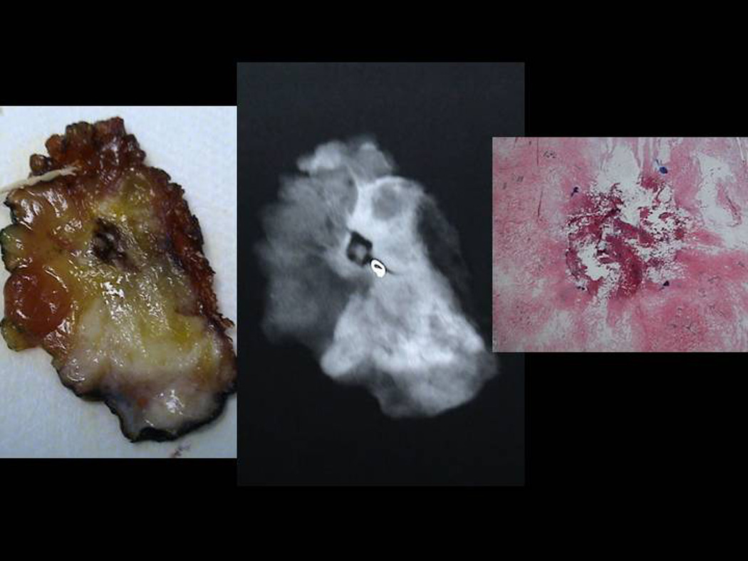
Figure 5.


Sectioned Pathology: a. 3mm serial sections of the lumpectomy specimen post PeRFA b. Xray demonstrating clip in biopsy cavity. c. Magnified view of biopsy cavity. d. Magnified view of xray of biopsy cavity demonstrating clip. e. Pathology PeRFA cavity site demonstrating no residual tumor and pericavitary ablation.
