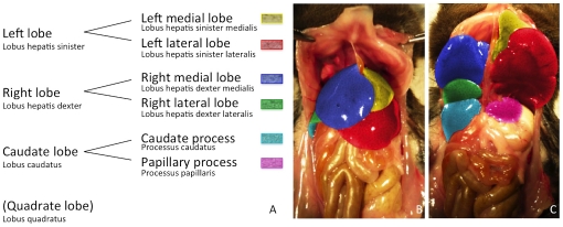
Figure 1. Illustrating the segmentation of the murine liver lobes according to the Nomina Anatomica Veterinaria.
A Since in most animals we have not been able to identify the quadrate lobe in micro-CT imaging or in situ, the quadrate lobe was put in brackets and not color-coded. B and C are photographs of the liver of a C57BL/6J mouse (ventral view). In C the left and the right liver lobe have been folded cranially to reveal the liver lobes lying below. The single liver lobes have been consistently color-coded according to the schematic segmentation (1A) and to the related micro-CT figures 2, 3, 4 to simplify orientation.

