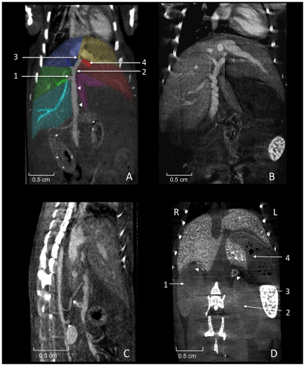
Figure 6. Perihepatic structures.
A shows coronally reconstructed micro-CT images acquired 20 minutes after injection of 400 µl Fenestra VC. The portal vein (arrowheads) runs in a caudocranial direction and divides into single veins supplying the liver lobes with blood from the abdominal organs. The right branch (1; Ramus dexter) divides up into two non-specified branches supplying the right lateral lobe (colored green) and the right caudate process (colored cyan). The left branch (2; Ramus sinister) divides into the umbillical part (3; Pars umbillicalis) and the transversal part (4; Pars transversalis) draining the left medial lobe and the left lateral lobe, respectively. The vein supplying the right medial liver lobe (colored blue) is not classified in the NAV. Spontaneous peristaltic movement of the portal vein is known to result in a corkscrew-like appearance of the vessel as shown in B. The liver is supplied with arterial blood from the coeliac artery (arrow) arising from the aorta cranial of the cranial mesenteric artery (arrowhead), as shown in C. The portal vein collecting blood from smaller abdominal vessels can be spotted directly ventral to the hepatic artery. D shows a coronally reconstructed micro-CT, 21 days after injection of 100 µl Exitron nano 12000 showing relevant organs adjacent to the liver. The right kidney (1), the left kidney (2), the spleen (3), the stomach (4; containing air bubbles), and the adrenal glands (arrow heads). The dotted lines indicate the renal impression of the liver on the right side and the gastric impression on the left side.

