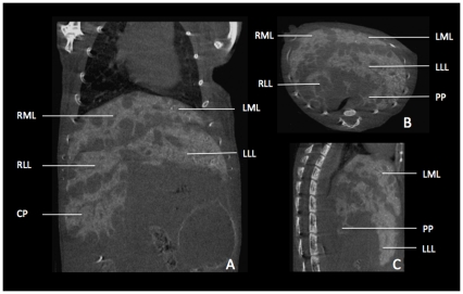
Figure 8. Illustrative micro-CT images of liver metatstases in a mouse.
A–C Images of liver metastases were acquired 2 hours after i.v. injection of 100 µl of a contrast agent (Viscover Exitron nano 6000; Miltenyi Biotec, Bergisch-Gladbach, Germany) in a coronal (A), transversal (B), and saggital (C) slice orientation. With regard to liver anatomy, we found metastases to develop predominantly under the liver capsule adjacent to the fissures between the liver lobes. While the left lateral lobe (LLL) shows no metastases on its caudal edge in this slice, there are plenty on its cranial edge, next to the left medial lobe (LML). The lobes on the right side (right medial lobe (RML), right lateral lobe (RLL) and the caudate process (CP)) show (besides the subcapsular growth pattern) metastases within the liver parenchyma. Due to tumor growth the papillary process depicted in B and C lost its typical shape (compare with figs. 2, 3, 4).

