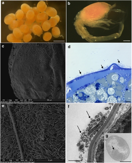
Figure 1.
Microscopic observations of eggs and larvae (SEM, c and e; TEM, f and g). (a) R. exoculata eggs from Logachev. Scale bar=500 μm. (b) R. exoculata larvae, which just hatched from Logachev (manually separated from egg). Scale bar=200 μm. (c) Egg surface from Logachev covered by thin rod-shaped bacterial mat. (d) Egg thin section from Logachev, showing bacterial mat (indicated by dark arrows) on R. exoculata egg membrane. Scale bar=10 μm. (e) Magnified view of the picture in panel c, showing thin rod-shaped bacterial mat. (f) Egg thin section, showing thin rod-shaped bacteria (indicated by dark arrows) on the egg membrane. Scale bar=2 μm. (g) Methanotrophic-like bacteria (with intracytoplasmic membranes, indicated by a dark arrow) retrieved in the thin rod-shaped bacterial mat associated with the egg membrane. Scale bar=500 nm.

