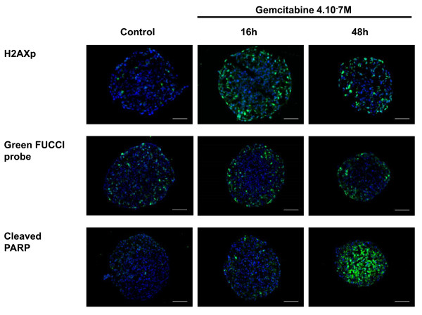
Figure 4.
Spatio-temporal response of Capan-2 spheroid to gemcitabine. Analysis of gemcitabine response was done on 5 μm frozen sections of Capan-2 spheroids treated 16 h and 48 h with 4.10-7 M gemcitabine. DNA damage was revealed by immunodetection of phosphorylated of γH2AX, S phase checkpoint was monitored on Capan-2 spheroid expressing the geminin-mAG FUCCi green probe and apoptosis was analyzed by immunodetection of cleaved form of PARP. Images were collected using a X10 objective. The scale bar corresponds to 100 μm. Results shown are representative of the examination of 3 sections from 5 spheroids. Each experiment has been repeated 3 times.

