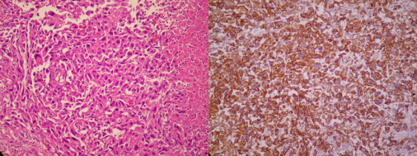
Figure 2.

The pathological features of hepatic angiosarcoma (Case 9). The sections of the specimen show a picture of liver tissue with sheets of spindle tumor cells with focal necrosis (Left, H&E stain, x200). Focal vascular channels are also lined by atypical cells. The tumor cells show positive staining in the CD31 (Right, x200), CD34, factor VIII, D2-40 and c-kit stains, and negative for AE1/AE3, hepatocyte, S100, and HHF-35 stains.
