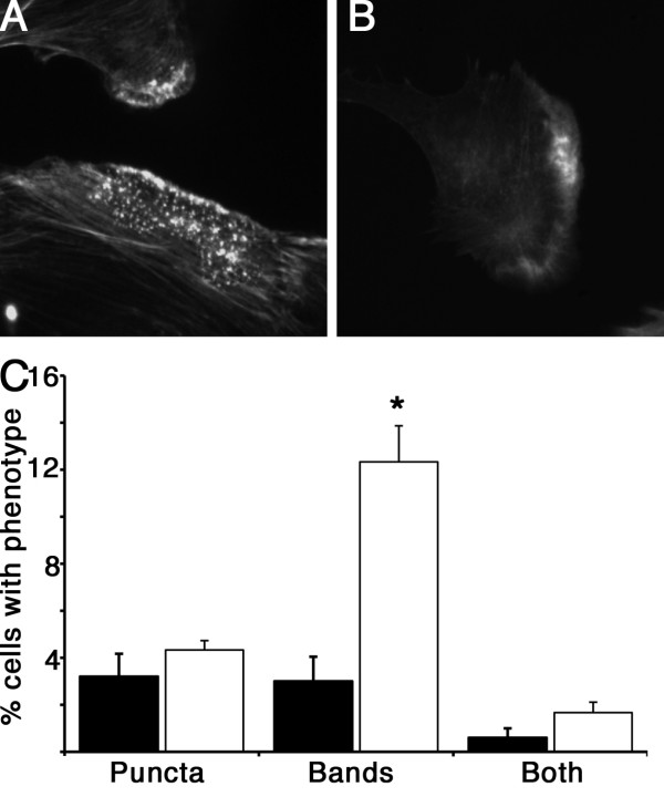
Figure 5.
REF52 cells depleted for SM22 have an altered actin morphology. SM22 depleted cell show an increase in dense actin puncta reminiscent of podosomes (A), and cells with peripheral actin bands (B), or both, but only the increase in peripheral actin bands was statistically significant. Quantitative data shown in C, represent at least 100 cells counted in each of 5 independent experiments, mean ± SEM, * p < 0.05 compared to control shRNA. Black bars, control shRNA, white bars knockdown clone 1B.

