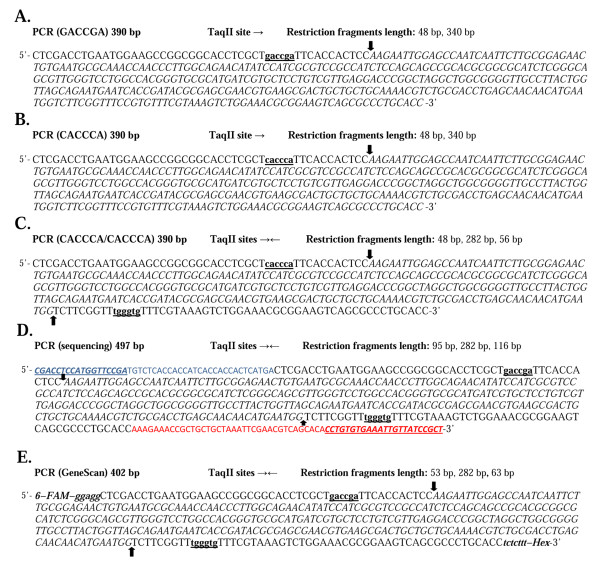
Figure 1.
PCR fragment DNA substrates. Putative recognition sequences of TaqII are in bold and underlined. Arrows mark the cleavage. The restriction fragments lacking TaqII recognition sequence are in italics. (A) PCR DNA fragment with a single 5'-GACCGA-3' site. (B) PCR DNA fragment with a single 5'-CACCCA-3' site. (C) PCR DNA fragment with two 5'-CACCCA-3' sites. (D) PCR DNA fragment used for sequencing reactions. The distances from the 5'- and 3'-ends to the TaqII recognition sites were extended using an additional pair of primers. The introduced DNA fragments are shown in blue for the forward primer and in red for the reverse primer. (E) PCR DNA fragment used for GeneScan analysis. 6-FAM and Hex denotes the fluorescence dyes. The introduced DNA fragments are shown in bold and italics.

