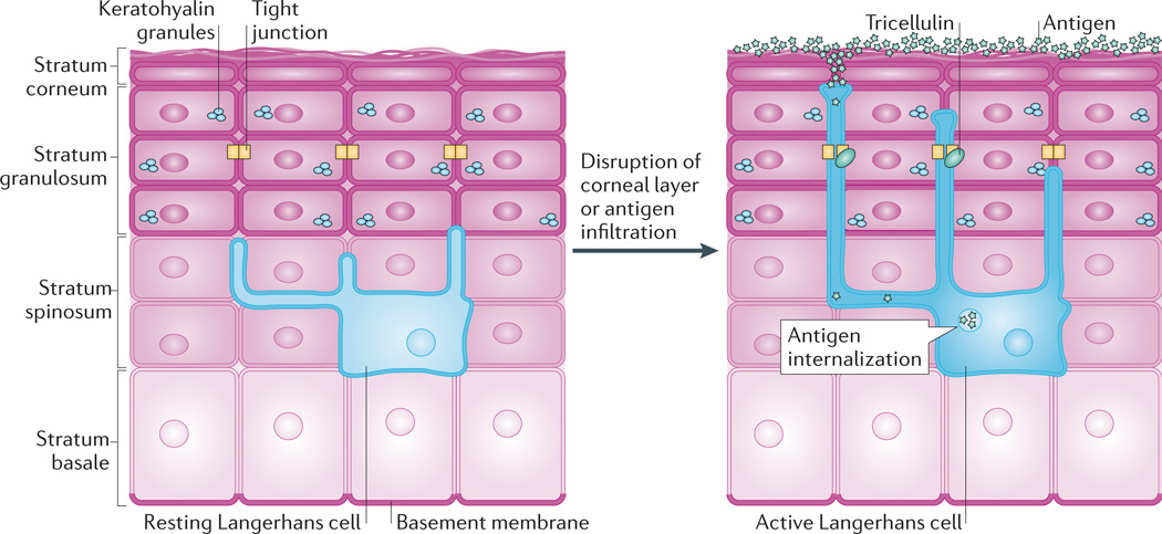
Figure 5. Tight junction dynamics during antigen sampling.
Tight junctions contribute to the barrier between the superficial epidermis and underlying stratum spinosum but must also be dynamic to allow antigen sampling. In the resting state, Langerhans cells reside among keratinocytes in the stratum spinosum and extend dendrites through suprabasal layers. When activated, for instance by disruption of the stratum corneum or by infiltration of antigens, the dendrites of Langerhans cells dock with tight junctions and gain the ability to extend into the stratum corneum. The transmembrane protein tricellulin localizes to these tricellular tight junctions formed between keratinocytes and Langerhans cells. This dynamic adjustment of tight junctions allows Langerhans cells to internalize antigens, which can then be presented to the host immune system144.

