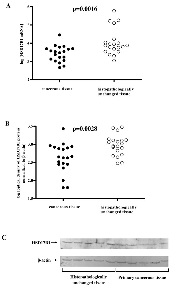
Figure 1.
HSD17B1 transcript (A) and protein (B) levels and representative picture of western blot (C) in primary cancerous and histopathologically unchanged tissues from patients with CRC in the proximal colon. The cancerous (●) and histopathologically unchanged tissues (○) from twenty patients with CRC in the proximal colon were used for RNA and protein isolation. Total RNA was reverse-transcribed, and cDNAs were investigated by RQ-PCR relative quantification analysis. The HSD17B1 mRNA levels were corrected by the geometric mean of PBGD and hMRPL19 cDNA levels. The amounts of HSD17B1 mRNA were expressed as the decimal logarithm of multiples of these cDNA copies in the calibrator. Proteins were separated by 12% SDS-PAGE, and transferred to a membrane that was then immunoblotted with Gp anti- HSD17B1 Ab and incubated with donkey anti-goat HRP-conjugated Ab. The membrane was then reblotted with anti-actin HRP-conjugated Ab. The amount of western blot-detected HSD17B1 proteins was presented as the decimal logarithm of HSD17B1 to β-actin band optical density ratio. The p value was evaluated by unpaired, two-tailed t-test.

