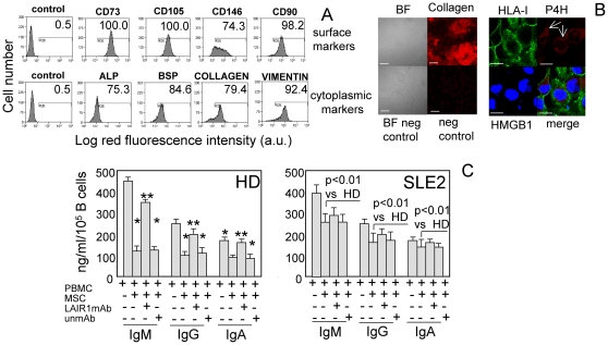
Figure 4. Collagen-producing LMSC regulate Ig production upon engagement of LAIR1: this effect is defective in SLE patients.
A, LMSC from a representative reactive lymph node were surface stained with the indicated mAbs (first row) followed by PE-conjugated anti-isotype specific goat anti-mouse antiserum. Control: cells stained with an unrelated mAb followed by GAM as negative control. Second row: LMSC were cytoplasmic stained after fixation and permeabilization with mAbs to the indicated molecules (ALP: alkaline phosphatase, BSP: bone sialoprotein, collagen or vimentin) followed by PE-conjugated GAM. Results are expressed as log red fluorescence intensity vs number of cells. In each panel are indicated the percentages of positive cells above the horizontal bar set on negative control (first subpanel on the left of each row). B. left: bright field (BF) of LMSC from a representative reactive lymph node (upper left), staining with anti-collagen mAb (upper right, red) without cell permeabilization and the respective negative controls (BF neg control, neg control). B right: staining of LMSC with anti-HLA-I (surface, green), anti-prolyl-4-hydroxylase (P4H, cytoplasmic, red) and anti-HMGB1 mAb (nucleus, blue) analyzed by confocal microscopy. Merge analysis is also shown. 400× (left), 600× (right) magnification. White Bars: 10 µ m; reactivity for collagen is disposed in large and concentrated regions (upper left); the white arrows indicate the intracytoplasmic reactivity for P4H (upper right). C. PBMC of healthy donors (HD, n = 7, left) or SLE patients (n = 9 from SLE2 group, right) were incubated for 5 d on collagen-producing mesenchymal stromal cells (MSC) from reactive lymph node coated plates. Then SN were analyzed for the presence of the human IgM, IgG and IgA by ELISA. In some experiments, F(ab′)2 of anti-LAIR1 mAb (5 µ g/ml) to compete with the interaction of surface LAIR1 and collagen-producing MSC or an unrelated mAb matched for the isotype as control mAb (5 µ g/ml) was added at the onset of cell culture. Results are expressed as ng/ml/105 CD20+ B cells as mean±SD. * p<0.001 vs basal production of Ig. ** p<0.001 vs Ig production on LMSC coated plates. In the right panel is indicated the statistical significance of Ig production in the culture condition PBMC+LMSC in SLE2 patients vs HD.

