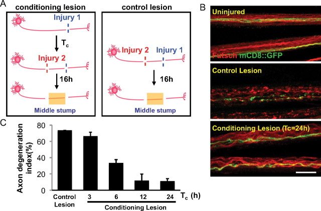
Figure 3.
A conditioning lesion protects axons from degeneration after a second injury. A, Schematic of the conditioning lesion and control lesion experiments. A conditioning lesion was done at injury site 1 (see Materials and Methods) for a variable period of time, Tc, before the second injury at site 2, which was within the proximal stump of the previously injured axon. B, Uninjured axons or middle stump axons in control and conditioning lesion were stained for Futsch (red) and GFP (green). Single axons, labeled by driving UAS-mCD8::GFP expression with m12-Gal4, and endogenous Futsch were fragmented at 16 h after the control lesion, but intact after the conditioning lesion. Scale bar, 25 μm. C, Quantification of the axon degeneration index, measured at 16 h after the second injury. For the conditioning lesion, the time between the first and second injuries, Tc, was varied.

