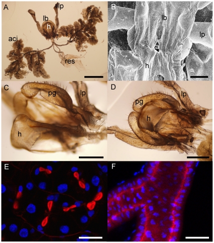
Figure 3. Raspy cricket labial glands.
(a) Labial glands and labium of Hyalogryllacris; scale bar is 2 mm. (b) Scanning electron micrograph of a preparation from Apotrechus in which the hypopharynx has been pinned back to show the aperture through which the labial glands empty into the salivarium; scale bar is 500 µm. (c) Labium and hypopharynx in open position, showing the raised margins on the labial paraglossae of Hyalogryllacris; scale bar is 500 µm. (d) Same preparation as (c), labium and hypopharynx in closed position, with raised margins of paraglossae overlapping hypopharynx; scale bar is 500 µm. (e) Confocal slice through labial acinus of Apotrechus showing paired arrangement of nuclei (blue) and secretory invaginations (red); scale bar is 50 µm. (f) Projection of 18 images in z-series showing small cuticle-secreting cells lining the acinar labial duct of Apotrechus; scale bar is 50 µm. aci = acini; res = reservoirs; lb = labium; h = hypopharynx; lp = labial palp; a = aperture of labial glands into salivarium; pg = paraglossae of labium.

