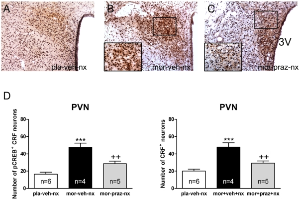
Figure 4. Increased pCREB into CRF neurons after naloxone-induced morphine withdrawal is α-1 adrenoceptor dependent.
PVN tissue isolated from placebo or morphine-dependent rats pretreated with vehicle or prazosin before naloxone injection was processed for pCREB and CRF double-label immunohistochemistry. Top panels (A–C) represent immunohistochemical detection of pCREB into CRF neurons after the different treatments. Low and high magnifications images show pCREB-positive (blue-black)/CRF-positive (brown) neurons in the PVN. Scale bar: 100 µm (low magnification); 20 µm (high magnification). 3V, third ventricle. Bottom panels (D) show quantitative analysis of pCREB-positive/CRF-positive and total CRF-positive (with or without pCREB) neurons in the PVN. Data shown are means ± SEM. Post hoc test revealed a higher number of pCREB-positive nuclei in CRF immunoreactive neurons after naloxone-induced morphine withdrawal. This increase was antagonized in prazosin-pretreated rats. The increase in number of CRF-positive neurons during morphine withdrawal was also blocked by prazosin. ***p<0.001 versus placebo (pla)+vehicle (veh)+naloxone (nx); ++p<0.01 versus mor+veh+nx.

