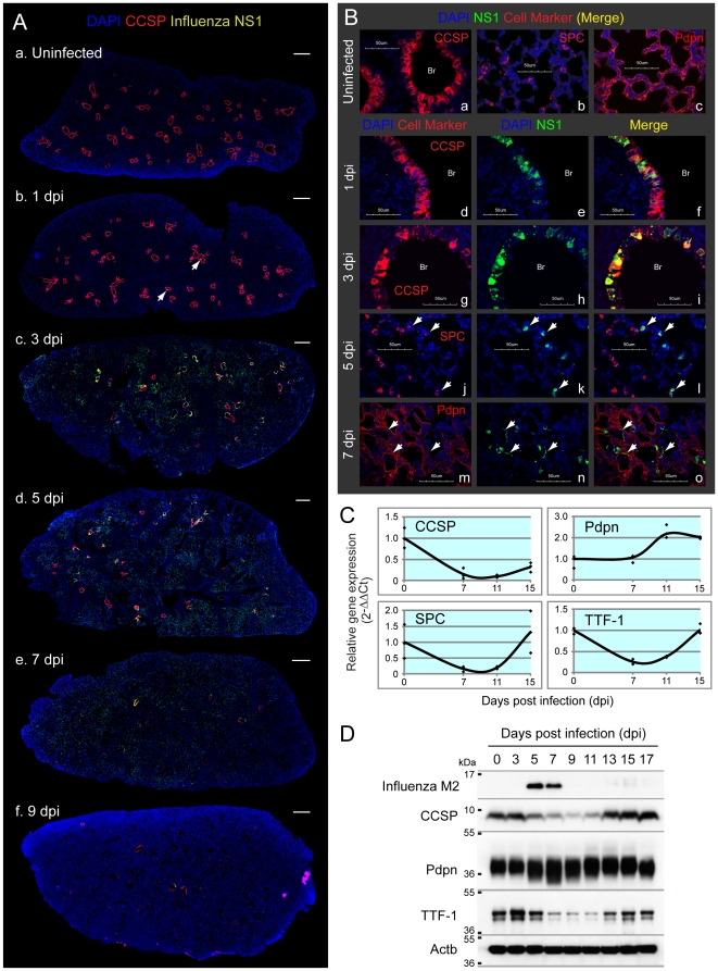
Figure 3. Clara and AT2, but not AT1 cells, are the major PR8-permissive cell types in mouse lung.
Mice were infected with sub-lethal dose of PR8 by intra-tracheal inhalation. A. PR8 replication sites (viral nonstructural protein 1 [NS1]-positive cells) spread from bronchial cells to alveoli with the peak at 5 dpi and almost completely disappeared by 9 dpi. Lung sections were immunostained with CCSP, NS1 and Prdx6. Slides were scanned by MIRAX MIDI system with the exact same exposure times for DAPI, CCSP, Prdx6 and NS1, respectively, and images were shown with the same adjustment of brightness and contrast. Scanned image for Prdx6 was shown in Fig. 5A. The scale bars represent 500 µm. Infected cells at 1 dpi were pointed with arrows. B. NS1 was mainly detected in bronchial (Br) Clara cells at 1 dpi (d, e, f), and then a layer of infected Clara cells lysed at 3 dpi (g, h, i). Alveolar infected cells at 5 dpi were co-stained with SPC (white arrows in j, k, l). NS1-positive AT1 cells (white arrows in m, n, o) were sometimes observed at mildly inflamed area at 7 dpi. Lung sections were immunostained with the indicated antibodies and observed at ×60 magnification with confocal microscope. The scale bars represent 50 µm. C. Gene expression of CCSP, SPC and TTF-1, but not Pdpn, was reduced following infection. mRNA expression in the lung tissue was investigated by qRT-PCR. Beta-actin was used for normalization. D. PR8 infection reduced protein levels of CCSP and TTF-1, but not Pdpn. Lung protein extracts from three mice were pooled, and were subjected to immunoblotting analysis with the indicated antibodies.

