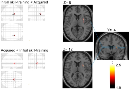
Figure 5. 11C-raclopride BP changes in sub-regions of the striatum between the two conditions.
Left panel shows the grass brain map of the voxel-based analysis of 11C-raclopride BP change (upper figure: initial skill-training condition < acquired condition and lower figure: acquired condition < initial skill-training condition). Right panel shows coronal and axial sections of the statistical parametric map of 11C-raclopride BP change in the initial skill-training condition versus acquired condition overlaying the MRI T1 image in stereotaxic space. Right side image corresponds to right side brain. The displayed cluster shows the significant area of decreased 11C-raclopride BP in the right antero-dorsal and lateral part of the putamen. The peak coordinate in the right putamen was located at X = 30, Y = 4, Z = 12. No BP change was observed in the left putamen.

