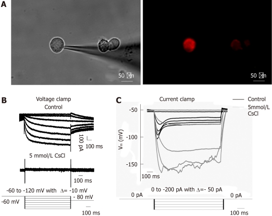
Figure 3.
Ih recorded under whole-cell voltage and current clamp from DiI labeled dissociated dorsal root ganglia and nodose ganglia neurons innervating the jejunum. A: DiI labeled dissociated dorsal root ganglia neurons; B: Representative current traces of Ih current (top trace); Current recorded in the presence of 5 mmol/L CsCl (middle trace); voltage clamp protocal: Ih was induced from a holding potential of -60 mV in 1 s pulses from -60 mV to -120 mV in steps of 10 mV, followed by a final step to -80 mV to record the tail current (bottom trace); C: Voltage response to test current pulse before (black) and after (grey) application of 5 mmol/L CsCl (top trace); bottom trace shows the current clamp protocol, i.e., hyperpolarizing current pulses ranging from 0 pA to -200 pA in steps of 50 pA. Note that the current elicited an instantaneous hyperpolarization that was followed by depolarization (named “sag”) of membrane potential. DRG: Dorsal root ganglia; NG: Nodose ganglia.

