
Figure 1.
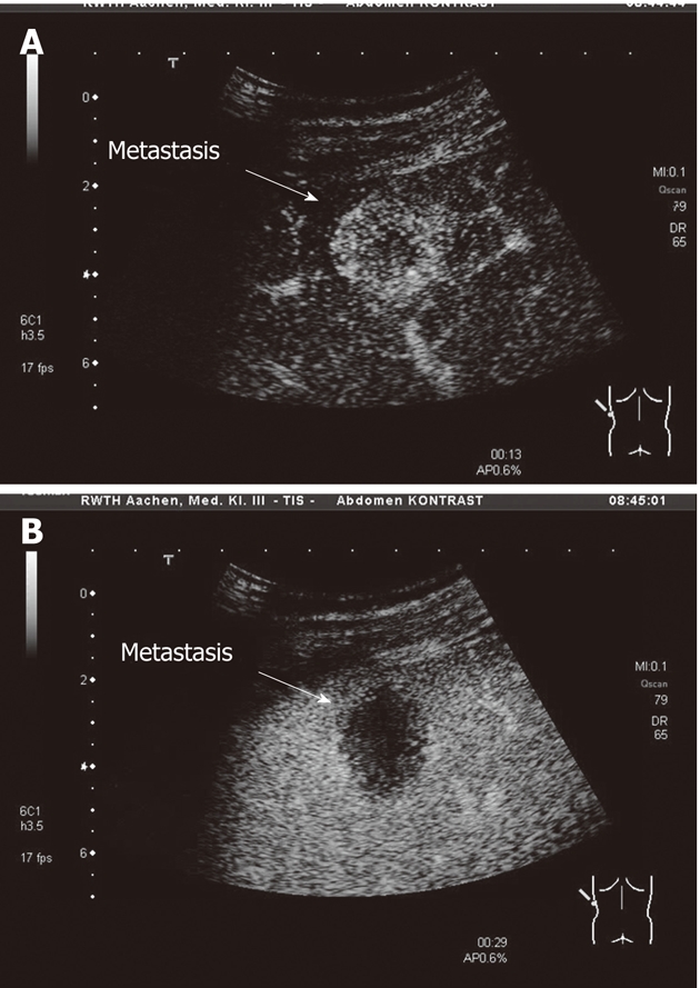
Contrast enhanced ultrasound. A: A representative liver metastasis after 13 s of contrast agent injection with early contrast enhancement; B: A representative liver metastasis after 29 s of contrast agent injection with lost of contrast enhancement.