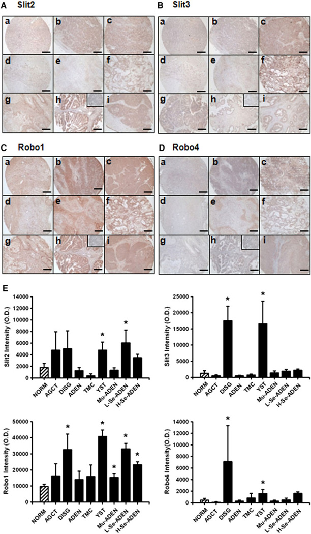
Fig. 1.
Immunohistochemical analysis of Slit2/3 and Robo1/4 in human ovarian cancer tissue microarray. Immunolocalization of Slit2 (A), Slit3 (B), Robo1 (C), and Robo4 (D) was performed as described in “Materials and Methods”. Reddish color indicates positive Slit and Robo staining. For each target protein, representative images from NORM (a), AGCT (b), DISG (c), ADEN (d), TMC (e), YST (f), Mu-ADEN (g), L-Se-ADEN (h), and H-Se-ADEN (i) are shown. For the goat (A, B) or rabbit (C, D) preimmune IgG control, representative images from the L-Se-ADEN are shown. Bar 200 µm. For the semiquantitative analysis of the Slit/Robo staining intensity (E), data are expressed as means ± SEM of the integrated OD. *Differs from NORM (p ≤ 0.05)

