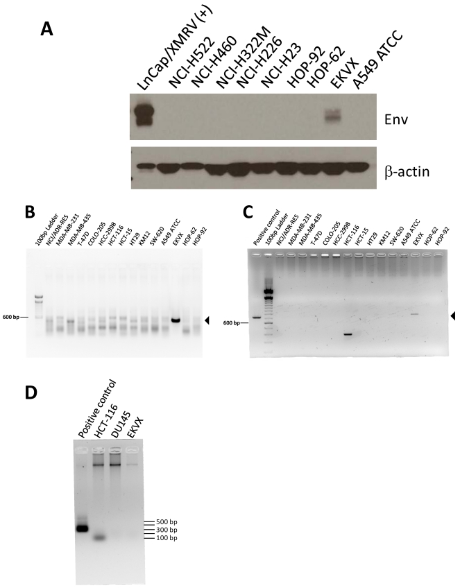
Figure 1.
Detection of viral protein and DNA in the human lung adenocarcinoma cell line EKVX. Immunoblotting and PCR were carried out on all 60 cell lines of the panel; a subset are shown. Immunoblotting of total protein lysates from lung cancer cell lines for Env with monoclonal antibody 7C10 (A). XMRV-infected LNCaP cell lysate [LNCaP/XMRV(+)] is included as a positive control. Env is present as both a precursor form (upper band) and a processed surface unit (lower band). Immunoblotting with β-actin antibody was used to confirm equal loading. Single-round PCR of genomic DNA for env (B) and gag (C) sequences. Template is genomic DNA from cell lines of the NCI-60 panel. Arrowheads indicate the expected fragment sizes: 533 bp for env and 731 bp for gag. Negative (no template) controls were run on separate gels and no products of the expected size were observed (data not shown). (D) EKVX and other representative cell lines, DU145 and HCT-116, were tested for mouse contamination by PCR for IAP using 600 ng genomic DNA as template. The positive control is genomic DNA from a mouse cell line diluted 1/105 in human cell line genomic DNA.

