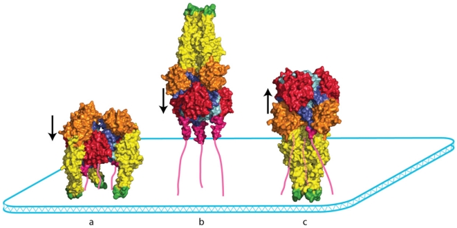
Figure 4.
The topological problem associated with VSV G conformational changes. The VSV G pre- and post-fusion conformations are represented by a space-filling model viewed from the side. The C-terminal segments that reach the membrane are depicted as pink lines. In the pre-fusion conformation (a) and in a putative trimeric intermediate (b), the three fusion domains are located outside a pyramidal volume where the base would be the membrane and the sides would be defined by the ectodomain C-terminal segments. The converse occurs in the post-fusion state (c). Going from the pre-fusion trimer to the post‑fusion trimer is very complicated, if not impossible, without monomerization. The arrows indicate the orientation of the central helices in each conformation.

