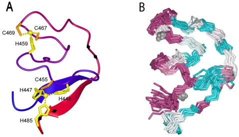
Figure 3.
NMR structure of the zinc-binding domain in the cytoplasmic tail of G2. (A) Ribbon model of the deduced structure. Coloring changes from blue (N terminus) to red (C terminus). The zinc ions are depicted as spheres, and the coordinating cysteine and histidine sidechains are displayed. Black diamonds on the C-terminal half of the ribbon depict the two pairs of basic residues (KK and RR) implicated in ER retention/retrieval. (B) Superimposed backbone traces are colored to indicate the degree of sequence conservation among arenaviruses: highly conserved residues are shown in purple and highly variable residues in cyan.

