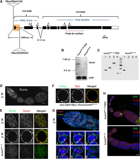
Figure 1.
Kumo localizes to the nuage and nucleus in the germarium and is required for oocyte fate maintenance. (A) Schematic representation of the kumo locus, consisting of two previously annotated genes, CG14303 and CG14306, which are 51.6 kb apart. The region highlighted in yellow represents a deletion of 460 bases in the kumoM41-13 allele by FRT recombination, which encompasses the first exon and the following intron, including the predicted start codon. Positions of PiggyBac insertions are represented in CG14306. The arrows indicate the primer sets used for RT–PCR. (B) Northern blot analysis of the kumo transcript. A transcript of ∼6 kb was detected in the ovarian RNA extracted from the control, but it was not detected in that from kumoe03728 ovaries. (C) RT–PCR with the primer sets denoted in Figure 1A showing the absence of the kumo transcript in kumoM41-13 ovaries. Lane 1, actin control; lanes 2–4, primers against either CG14306 or CG14303 as shown in Figure 1A. (D) y w ovariole immunostained for Kumo showing the expression in germline cells. Perinuclear foci in nurse cells are discernible. Scale bar: 20 μm. (E) (Upper panel) Co-localization of Kumo (red) with a known nuage component, Krimp (green). Scale bar: 5 μm. (Middle panel) Closer view of a single nurse cell nucleus. (Lower panel) Kumo expression is undetectable in a kumoM41-13 egg chamber, and Krimp was mislocalized from the perinuclear nuage. Scale bar: 5 μm. (F) A single nurse cell nucleus showing the co-localization of Myc–Kumo (red) with Krimp (green). (G) (Upper panel) Nuclear localization of Kumo (red, indicated with arrow) in the germarium, which was co-stained for Lamin (green) and DAPI (blue). Scale bar: 10 μm. (Lower panels) Optical sections of germline cells in germarium showing the perinuclear and nuclear foci of Kumo. (H) The control heterozygous and kumoM41-13 ovarioles stained for oocyte markers Orb (green) and C(3)G (red) and DAPI (blue). Orb and C(3)G are undetectable in the egg chambers of stage two and onward in the kumoM41-13 ovary. Scale bar: 10 μm.

