Figure 2.
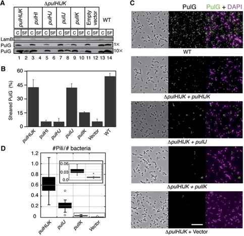
PulI and PulJ initiate pilus assembly in the ΔpulHIJK mutant. (A) PulG detection in cell and sheared fractions (C, SF) from WT and the ΔpulHIJK mutant complemented with empty vector or genes pulHIJK, pulHJ, pulHI, pulIJ and pulIK. The equivalent of 0.005 OD600 nm units (1 ×) or 0.05 OD600 nm units (10 ×) of C and SF was analysed. (B) Percentage of PulG in SF (mean+s.d.) from three independent experiments like the one shown in (C) with 0.05 OD600 nm equivalent analysed. (C) IF and phase contrast microscopy of E. coli expressing the pul genes (WT) on a plasmid or its minor pseudopilin deletion derivative (ΔpulHIJK). ΔpulHIJK mutant complemented with empty vector or genes pulHIJK, pulIJ and pulIK. PulG staining is in green and DAPI in magenta. Scale bar, 10 μm. (D) Box plot of normalized number of pili observed in (A) from ∼40 randomly selected areas in two independent experiments. Figure source data can be found in Supplementary data.

