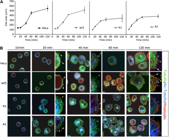
Figure 6.
Expression of GRK2 mutants defective in HDAC6 regulation results in an altered cell spreading pattern. (A, B) Parental, wt5, A1 or K1 HeLa cells were plated on coverslips coated with FN (10 μg/ml), fixed at the indicated times and analysed by confocal microscopy. The spreading area was quantified by morphometric analysis (A) and cells were triple stained (B) for acetylated α-Tubulin (blue), α-Tubulin (green) and F-actin (Phalloidin, red) as described in Materials and methods. Zoomed images are shown at 20, 40 and 120 min of spreading. Blue, green and white arrows and white arrowheads indicate acetylated MTs, non-acetylated MTs, pioneer MTs, and blebs, respectively.

