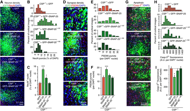
Figure 6.
SNAP-25 overexpression rescues neurodegeneration in CSPα KO mice. (A–C) GFP–SNAP-25 expression reverts neuron loss in CSP−/− mice. Lentiviruses expressing GFP alone, GFP–SNAP-25 WT, or truncated versions (GFP–SNAP-251−197 and GFP–SNAP-251−180) were injected into the cortex of littermate newborn CSP−/− mice. Mice were analysed at P50 by immunocytochemistry for NeuN puncta relative to DAPI puncta in the injected areas. (A) Representative merged images of GFP (green), NeuN (red), and DAPI signals (blue; for individual images, see Supplementary Figure S5). (B) Distribution of NeuN puncta density as percent of DAPI and (C) summary graph. (D–F) SNAP-25 overexpression rescues the reduction in synapse density in CSP−/− mouse cortex. (D) Following lentiviral expression of GFP alone, GFP–SNAP-25, GFP–SNAP-251−197, or GFP–SNAP-251−180, brain sections were immunostained for GFP (green), PSD95 (red), and DAPI (blue) at P50. PSD95 puncta were analysed as a function of DAPI puncta. (E) Distribution of PSD95 puncta as percent of DAPI and (F) summary graph. (G–I) SNAP-25 overexpression rescues the elevated apoptosis in CSP−/− mouse cortex. (G) Following lentiviral expression in vivo as above, the brain sections were immunostained for GFP (green), cleaved caspase-3 (Casp-3CL; red), and DAPI (blue) at P50. Total Casp-3CL positive pixels were analysed as a function of DAPI puncta. (H) Distribution of Casp-3CL staining as percent of DAPI and (I) summary graph. See Supplementary Figure S6A for individual images. Data shown are mean values±s.e.m.; n=3 mice, 10 sections for each; *P<0.05; ***P<0.001. Statistical significance was assessed by Student’s t-test, comparing each test condition to the control analysed in the same experiment.

