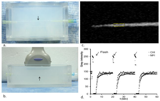
Fig. 1.
In vitro vessel phantom study. (a) Top view of a wall-less vessel phantom (arrow) made with 2% agarose gel. (b) US probe placement longitudinally to the wall-less vessel phantom (arrow). (c) Contrast harmonic image (CHI) of the phantom with microbubble solution flowing through. (d) Time- intensity curves of CHI and MFI with destruction of microbubbles (flash, arrow) every 20 seconds.

