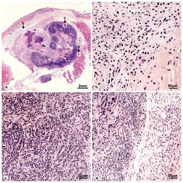
Fig. 6.
Histopathologic images (H&E stain) of a representative tumor on day 7 after RF ablation. A coagulation region (dotted line) locates at the side of the tumor. Central necrosis is visible in the residual viable tumor. Graphs b, c, d show the inflammatory rim of ablated region, residual viable tumor and residual tumor rim, respectively. Granulation tissue with inflammatory vessels is shown in inflammatory rim of ablated region (b). Extensive lymphocyte infiltrate is visible in the fibrous capsule of residual viable tumor with small vessels (d).

