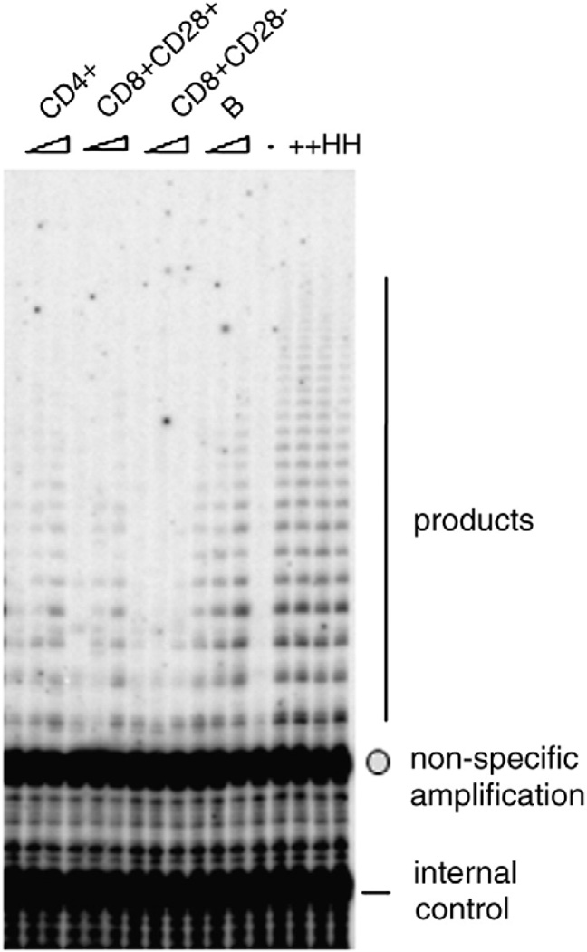
Fig. 1.
A typical gel phosphorimager image showing TRAP activity using the TRAPeze kit, from the four cell types from one study subject. +: Standard sample containing extracts from 20 293T cells. 2000, 5000 and 10,000 cells were used for each sample. The arrow points to the non-specific band and the circle points to the internal control band (see Materials and methods).

