Fig. 1.
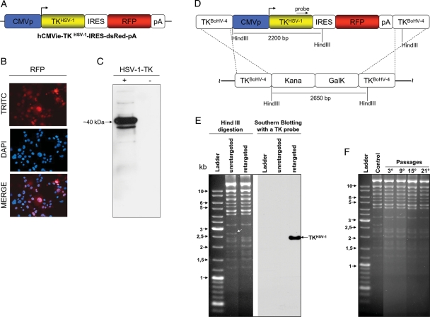
A, hCMVie-TKHSV-1-IRES-dsRed-pA expression cassette diagram (not on scale) containing the human cytomegalovirus immediate early promoter (CMVp; blue), the HSV-1 -TK ORF (TKHSV-1; yellow), an internal ribosomal entry site (IRES; white), the red fluorescent protein ORF (RFP; red) and a polyadenylation signal (pA; white). B, Representative image of cells transiently transfected with hCMVie-TKHSV-1-IRES-dsRed-pA, expressing the RFP as visualized by a fluorescence microscope with a red filter (TRITC) and their nuclei counterstained with DAPI, which co-localized with red cells when overimposed (MERGE). C, Western immunoblotting of the same hCMVie-TKHSV-1-IRES-dsRed-pA transiently transfected cells expressing HSV-1-TK (+). A negative control was performed with cells transiently transfected with an empty vector (-). D, Schematic diagram (not on scale) of the retargeting strategy employed on BAC-BoHV-4-A genome targeted at the TK locus with a KanaGalK selectable cassette (pBAC-BoHV-4-A-TK-KanaGalK-TK). The Kana/GalK cassettes were removed via heat-inducible homologous recombination and replaced with hCMVie-TKHSV-1-IRES-dsRed-pA expression cassette. E, The selected colonies were tested through HindIII restriction enzyme analysis, agar gel electrophoresis and Southern blotting. The retargeted clones were detected through the disappearance of the 2.6 kb band (indicated by a white arrow) and the appearance of a 2.2 kb band detected by Southern blotting. F, pBAC-BoHV-4-A-hCMVie-TKHSV-1-IRES-dsRed-pA clonal stability in Escherichia coli SW102 cells, passaged for 21 consecutive days and analyzed by HindIII digestion and agarose gel electrophoresis. The unretargeted pBAC-BoHV-4-A-TK-KanaGalK-TK was used as a control.

