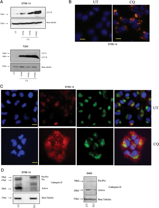
Fig. 3.
Chloroquine (CQ)–induced lysosomal stress blocks autophagy completion in malignant peripheral nerve sheath tumor (MPNST) cells. (A) CQ -treated cells (50 µM) demonstrated a time-dependent increase in levels of LC3II, as assessed by immunoblot anlaysis. (B) Cells exposed to CQ (25 µM; 24 h) demonstrated significantly higher levels of autophagic vacuole (AV)–associated LC3 immunoreactivity (red), relative to untreated (UT) cells. (C) UT cells showed discrete Lamp1-associated (green) cathepsin D immunoreactivity (red), whereas CQ–treated cells (50 µM; 24 h) exhibited diffuse cytoplasmic cathepsin D immunoreactivity. Cell nuclei were counterstained with bisbenzimide (blue). Scale bar equals 20 microns. (D) CQ (50 µM; 24 h) induced a decrease in levels of active cathepsin D, as assessed by immunoblot analysis.

