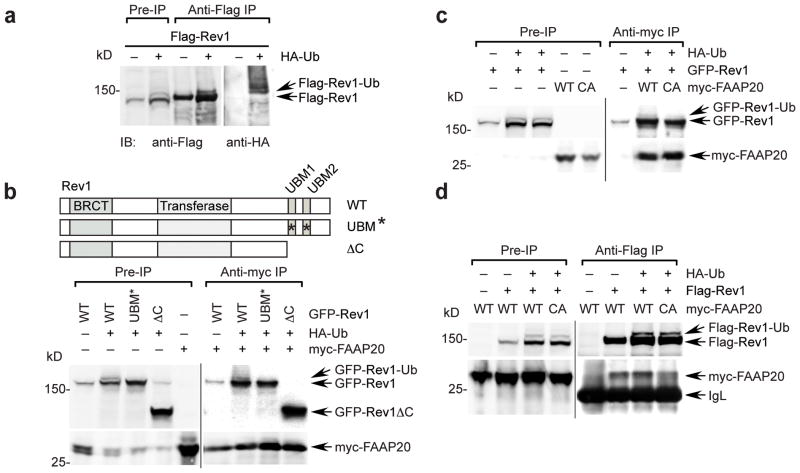
Figure 4. Monoubiquitination of Rev1 enhances the interaction with FAAP20.
(a) Monoubiquitin of Rev1 was visualized by anti-Flag immunoprecipitation and immunoblot of 293T cells transfected with cDNAs encoding Flag-tagged Rev1 with or without HA-tagged ubiquitin. The slower-migrating band, above Flag-Rev1, represents the monoubiquitinated form of Rev1, which was confirmed by anti-HA immunoblot. (b) (Top) schematic of Rev1 mutants. (Bottom) anti-myc immunoprecipitation of 293T cell lysates coexpressing HA-ubiquitin and GFP-tagged Rev1 wild-type, UBM* (L946A P947A L1024A P1025A), or ΔC (aa1–892) mutant, mixed with the lysates expressing myc-tagged FAAP20. (c) Anti-myc immunoprecipitation of 293T cell lysates coexpressing HA-ubiquitin and GFP-tagged Rev1, mixed with lysates expressing myc-tagged FAAP20 wild-type or CA mutant (d) Anti-Flag immunoprecipitation of 293T cell lysates coexpressing HA-ubiquitin and Flag-tagged Rev1, mixed with lysates expressing either myc-tagged FAAP20 wild-type or CA mutant.

