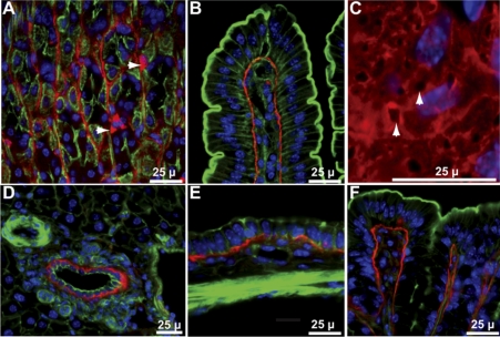
Fig. 2.
Gpx3 in tissues of Gpx3+/+ mouse gastrointestinal tract. A: stomach-arrowheads point to Gpx3-positive cells that have not been identified. B: duodenum. C: en face view of duodenal villus arrowheads point to basement membrane pores. D: liver. E: gall bladder. F: colon. Gpx3 is red; actin is green; nuclei are blue.

