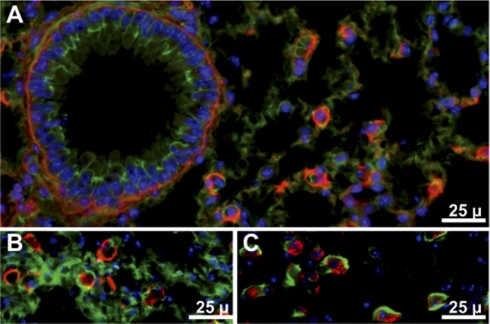
Fig. 4.
Gpx3 in mouse lung. In A and B Gpx3 is red, actin is green, and nuclei are blue. A: from Gpx3+/+ mouse. B: from Gpx3−/− mouse 2 wk after transplantation of a Gpx3+/+ kidney. C: from Gpx3+/+ mouse. In C, Gpx3 is green, antiprosurfactant protein C (stain for type II pneumocytes) is red, and nuclei are blue.

