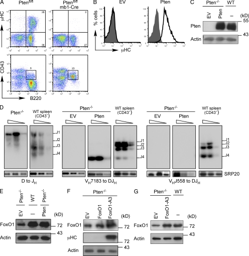
Figure 1.
Pten-deficient pro–B cells do not express µHC. (A) Flow cytometric analysis of BM cells from Ptenfl/fl mice and Ptenfl/fl/mb1-Cre mice (n > 3 per genotype), stained for the indicated surface markers. Numbers in quadrants indicate percentages of cells. (B) Flow cytometric analysis of µHC expression in Pten-deficient pro–B cells reconstituted with either an empty expression vector (EV) or with a Pten expression vector (black lines) as compared with untransduced cells (filled gray). (C) Immunoblot analysis of Pten expression in Pten-deficient pro–B cells transduced with an empty expression vector (EV) or with a Pten expression vector as compared with WT cells. Immunoblot of actin served as a loading control. (D) Southern blot analysis of PCR fragments amplified with specific primers for VHDQ52, VH7183, VHJ558, and JH4 from genomic DNA of Pten-deficient cells transduced with an empty expression vector (EV) or with a Pten expression vector. Wedges above lines indicate 2× serial dilutions. PCR for the splicing factor SRP20 was used as control. Purified CD43− splenic B cells from WT mice served as a control. (E) Immunoblot analysis of FoxO1 expression in Pten-deficient cells transduced with either an empty expression vector (EV) or a Pten expression vector as compared with WT cells. Immunoblot of actin served as a loading control. (F) Immunoblot analysis of FoxO1 and µHC expression in Pten-deficient cells transduced with either an empty expression vector (EV), a FoxO1 expression vector, or an expression vector encoding activated FoxO1 (FoxO1-A3). Immunoblot of actin served as a loading control. (G) Immunoblot analysis of Pten-deficient cells transduced with EV or FoxO1-A3 and analyzed for FoxO1 expression as compared with WT cells. Actin was used as a loading control. Data are representative of at least two to three independent experiments.

