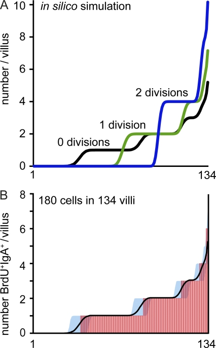
Figure 4.
Newly generated plasma cells are evenly distributed in the gut mucosa. (A) A random distribution of 180 cells throughout 134 individual villi was predicted by in silico simulation assuming no (black), one (green), or two (blue) cell divisions. (B) Mice received BrdU continuously with the drinking water for 7 d, and the number of BrdU+IgA+ cells in individual villi was enumerated by immunofluorescence microscopy. Inspecting 134 villi (pooled from five mice), we found 180 BrdU+IgA+ cells. The x axis corresponds to individual villi sorted from villi containing no cells to villi containing multiple BrdU+IgA+ cells, and each villus is represented by a vertical red line. Mean and 95% confidence interval of the in silico simulation with no cell divisions are depicted by a black line and blue-filled area. Similar results were observed after 2 d of BrdU application.

