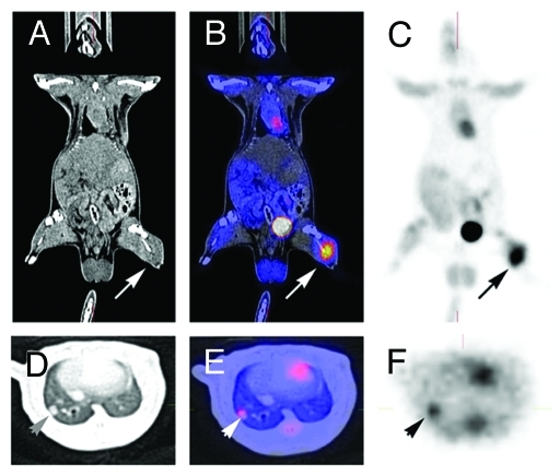
Figure 1.
PET-imaging and CT-scan leading to fusion images and allowing early detection of metastasis. A rat grafted with an osteosarcoma (no image from a metastatic breast cancer was available) in the left leg was explored by a by regular CT-Scan (A, D) by a PET-Scan technique (C, F). The images (B, E) were obtained by superposition of A and B, and D and F, respectively. A 3 mm metastasis (arrow) is detected in the lung both by CT-Scan and PET-imaging. Modern imaging techniques allow the early detection of metastasis, the measurement of metabolic activity of the primary cancer and of metastases, and the fusion of images obtained with different modalities. They allow the staging according to RECIST criteria.104

